Significance of PACS Medical Imaging in Modern Digital Radiology
Diagnosis and treatment are two essential elements; the medical system relies on to treat many diseases. A precise surgery couldn’t be possible without diagnosing the actual defect in an organ in the right location. Diagnosis and treatment have an integration because a proper diagnosis can help to choose the suitable treatment method. New diseases and many prevailing conditions affecting more people have created the significance of the invention of new ways of diagnosis.
Precise medical diagnosis technology
In an era of advanced digital technology, innovative digital radiology as a precise diagnosis technology has become a reality. Many innovative diagnostic methods have developed been in a past few decades, and they are successfully used in medical technology applications. Innovative digital technology techniques have offered an excellent solution to treat many rare and complex diseases that had high mortality in the past due to non-availability of right diagnosis. Physician, surgeons, and medical technicians nowadays make an in-depth study of the defects in their patient’ organs to arrive at the correct conclusion before the actual treatment.
Digital medical imaging technology
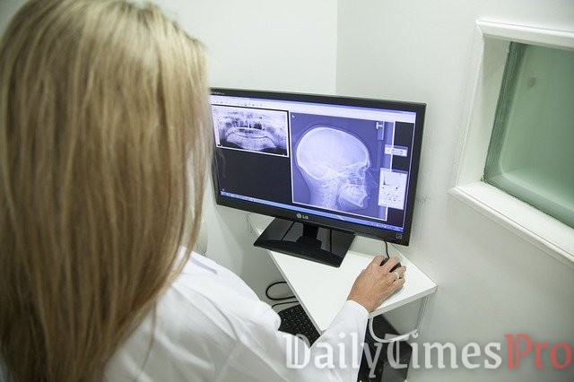
Digital medical imaging technology has revolutionized the field of medical diagnosis. Human anatomy is a complex integration of several organs and other elements, such as bones, muscles, arteries and veins, nerves, tissues, and many other minute structures that are quite hard to access precisely using a naked eye or conventional diagnostic methods. Digital technology uses extra-sensitive methods to capture crucial data needed during a patient’s examination directly. The collected data is instantly transferred to a computer system, where it is stored and used when it is required. A patient’s digital diagnostic data can also be transmitted to a medical practitioner or surgeon on any geographical location. Data can also be replicated to use of several places.
Significance of digital radiology systems
Digital medical imaging techniques applied in digital radiology are advanced diagnostic measures used for clinical analysis and medical intervention. They work by creating a database of normal physiology and anatomy for detecting abnormalities. They make visual representations of the body’s anatomy of organs and tissues meticulously, and to study their functions and physiology. Digital radiology systems are significant for the following reasons:
- They produce sharp 3D colored real-time images.
- Digital documents created as audio, stills, motion picture files, audios and videos can be analyzed correctly and quickly.
- Digital files can be transferred and stored on a computer system instantly after creation and accessed by the users in the medical profession online on any location across the globe.
- Original digital images remain intact and preserved for a more extended period due to reduced physical contact with the files.
- Digital files can also be printed for physical records.
How digital radiology methods work
Medical professionals and radiologists nowadays are trained to deal with advanced digital medical imaging techniques, as conventional medical diagnostic procedures like X-rays have become obsolete in detecting abnormalities correctly. They trust only on modern diagnostic methods to diagnose, monitor and treat medical conditions. The steps in the use of modern digital radiology systems include radiology reporting. It is a way of communication among healthcare professionals associated with the treatment of a particular patient for a specific medical condition for which certain clinical questions are required to be answered. The reporting is based on the set guidelines that require the inclusion of the complete medical history of a patient and its analysis by a competent radiologist.
The factors crucial factors to consider in the report are:
- Reason for a radiology exam
- Tests conducted under radiology exam
- A procedure used to conduct radiology exam
- Type of digital radiology systems used
- Outcomes of the investigation
- Radiologist’s analysis, findings (positive or negative), and comments on the result.
Computed radiography (CR) and Direct radiography (DR)
Digital radiology systems require computer networks and high-bandwidth web facilities. Two primary methods used nowadays are computed radiography (CR) and direct radiography (DR). The primary difference between the two technologies is that active matrix flat panels covered with a dynamic matrix array of thin-film transistors and photodiodes are used in DR systems, and CR cassettes use photo-stimulated luminescence screens for capturing the X-ray image. One cannot say which system out of DR and CR is the best because they are excellent for their specific applications. Magnetic Resonance Imaging (MRI), Radiographic Fluoroscopy (RF), and Ultrasound Imaging are examples of commonly used DR systems. CR systems are standard or high-definition (HD).
Benefits of PACS and its association with RIS
Most prevalent modern diagnostic medical imaging methods are echocardiography, elastography, functional near-infrared spectroscopy, magnetic resonance imaging (MRI), magnetic particle imaging, nuclear medicine, photoacoustic imaging, tomography, and ultrasound. PACS medical imaging is also one of the quite prevalent as a modern radiological system for medical diagnosis. Picture Archiving and Communication System (PACS) is a computerized replacement of conventional radiological film. The PACS is a very accurate and convenient method, as it stores the digital image and prepares radiological reports electronically. The system has four essential elements:
- Imaging modalities for actual scanning of a patient
- Secured database network for uploading and transmitting a scanned image
- A workstation that allows doctors and radiologists to access, study and analyze thew image
- Archives that is important for storage to maintain a secured record of the image. Archives provide availability of the image for authorized access by medical professionals.
PACS offers advantages, such as enhanced image visualization, better organization of patients’ data, and cost-effectiveness. Radiologist nowadays is keen on using PACS software for its advantages over conventional scanning. PACS software integrates with Radiology Information System (RIS) for better functioning of the radiology department functions in a hospital, as radiologists can use for recording radiology histories of patients and appointment scheduling. The images stored in PACS can be conveniently transferred to other departments quickly, wherever and whenever needed.
The use of PACS software is also beneficial for patients.
- PACS imaging reduce examination time considerably.
- The chance of re-capturing images is reduced in PACS.
- The high-quality imaging in PACS allows patients to receive a more precise diagnosis.
- Patients have a low risk of radiation exposure compared to X-rays and fewer side-effects.
Wrap up
Digital radiology systems are a breakthrough in the field of medical diagnostic for accuracy and effective treatment. PACS medical imaging is one of the best technologies today for many benefits.